
Buttock muscles, artwork Stock Image C020/0127 Science Photo Library
The buttocks ( SG: buttock) are two rounded portions of the exterior anatomy of most mammals, located on the posterior of the pelvic region. In humans, the buttocks are located between the lower back and the perineum.

4 Anatomy of the buttocks and pelvis (Image by Springer) Download Scientific Diagram
File usage on Commons The following 2 pages use this file: File:Surface Anatomy of Female Buttock.jpg File:Surface Anatomy of Female Buttock.png Metadata This file contains additional information such as Exif metadata which may have been added by the digital camera, scanner, or software program used to create or digitize it.
Gluteal region
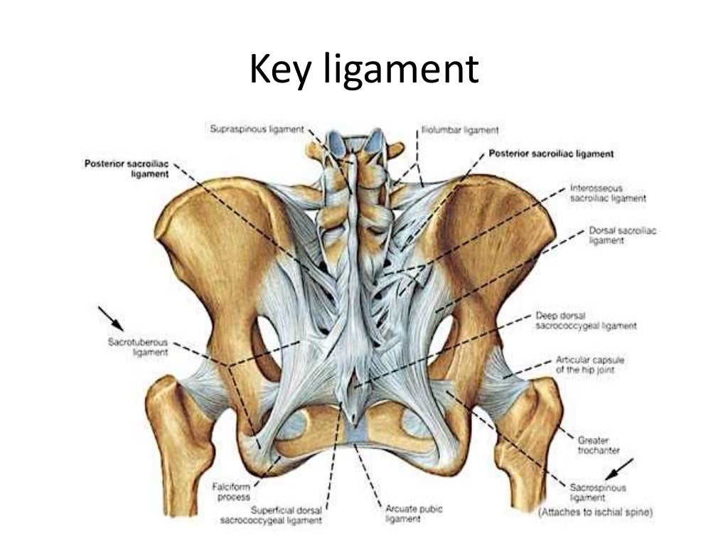
The hip joint The hip joint is a ball-and-socket joint, formed by the femoral head and the acetabulum (Fig. 1, see Standring, Fig. 80.15). The articular surfaces are spherical with a marked congruity; this limits the range of movement but contributes to the con-siderable stability of the joint.
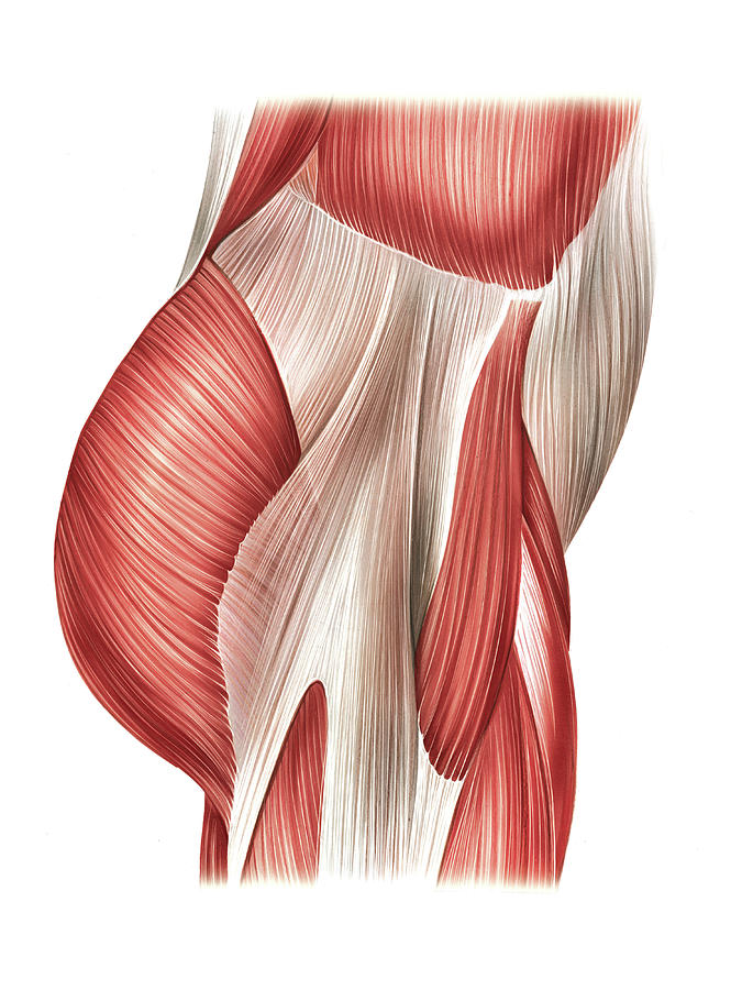
The Glorious Glutes Muscles of the Buttocks
Intergluteal cleft. The intergluteal cleft or just gluteal cleft, also known by a number of synonyms, including natal cleft, butt crack, ass crack and cluneal cleft, is the groove between the buttocks that runs from just below the sacrum to the perineum, [1] so named because it forms the visible border between the external rounded protrusions.

SOLUTION Medicine anatomy of the buttocks thigh and sacral plexus reviewer Studypool
I also searched Netter Images4 but did not find a detailed surface anatomy of the buttocks. Finally, I searched scholarly articles, using the CINAHL and MEDLINE databases. This search was based on the key terms "surface anatomy," "skin," "buttocks," and "gluteal area." This search did not retrieve useful visual aids. Creation of an Original Image

Buttock Muscles Anatomy Stock Photo 299396888 Shutterstock
The acetabular articular surface is an incomplete cartilaginous ring, thickest and broadest above, where the pressure of body weight falls in the erect posture, narrowest in the pubic region. The rough lower part of the cup, the acetabular notch, is not covered by cartilage.

posterior butt/thigh muscles Diagram Quizlet
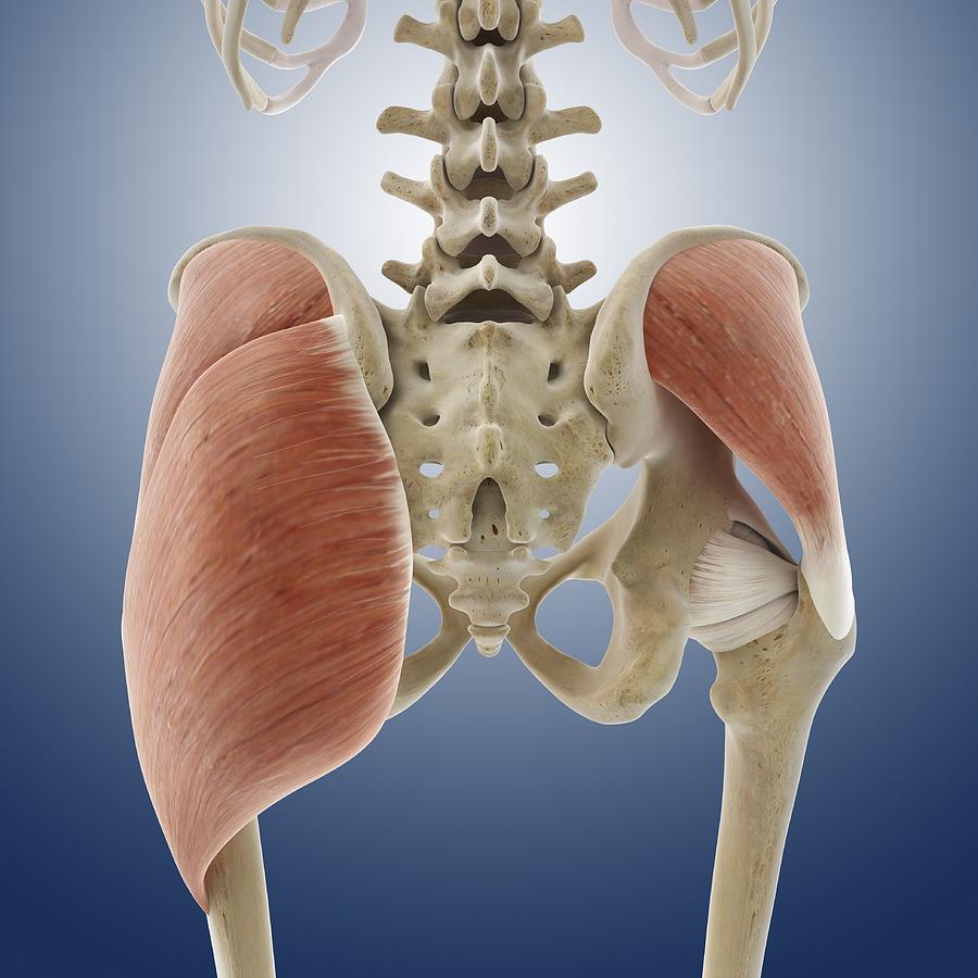
The gluteal muscles, also referred to as glutes or buttock muscles, are a muscle group consisting of the gluteus maximus, gluteus medius, gluteus minimus and tensor fasciae latae muscles. They are found in the gluteal, or buttock region, overlying the posterior aspect of the pelvic girdle and the proximal part of the femur.
Buttock muscles, artwork Photograph by Science Photo Library Pixels
Completing the comprehensive anatomical study, after general considerations, anatomical limits, surface anatomy, and descriptive anatomy, we will continue this chapter with the study of the surgical anatomy of the gluteal region, based on the dissection work of 20 cadavers performed by the author (Professor of Morphology at the Universidades.

Buttock muscles, artwork Stock Image C013/4419 Science Photo Library
Superficial abductors and extenders - group of large muscles that abduct and extend the femur. Includes the gluteus maximus, gluteus medius, gluteus minimus and tensor fascia lata. Deep lateral rotators - group of smaller muscles that mainly act to laterally rotate the femur.

Human buttock muscles, illustration Stock Image F012/7868 Science Photo Library
Bone Structure Muscles Subcutaneous Fat Skin Let's take a look at each of these in turn. Bone Structure The pelvis is the part of your skeleton which forms the shape of your hips and basis for your buttocks. This is one part of your butt that you cannot change.
Buttock Muscles Photograph by Asklepios Medical Atlas Pixels
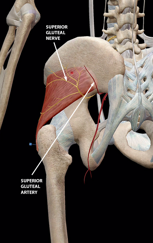
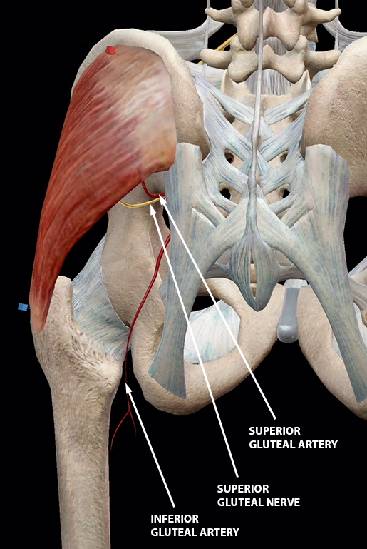
The gluteal region is an anatomically important area at the posterior aspect of the pelvis, which contains muscles critical to dynamic movements and upright stability of humans.[1][2] It is a key conduit for several important neurovascular structures passing into the lower limb.[3] The major muscles in this region are the gluteus maximus, medius, and minimus, of which the former is the largest.

Human buttock muscles, illustration Stock Image F011/6037 Science Photo Library
1/7 Synonyms: Anal cleft, Crena ani The intergluteal cleft is a surface anatomy landmark of the pelvis and lower limb. It is the deep furrow or groove that lies between the two gluteal regions (commonly known as the buttocks). It extends from sacral level S3 or S4 and ends just inferior to the apex of the sacrum, at the level of the anus.
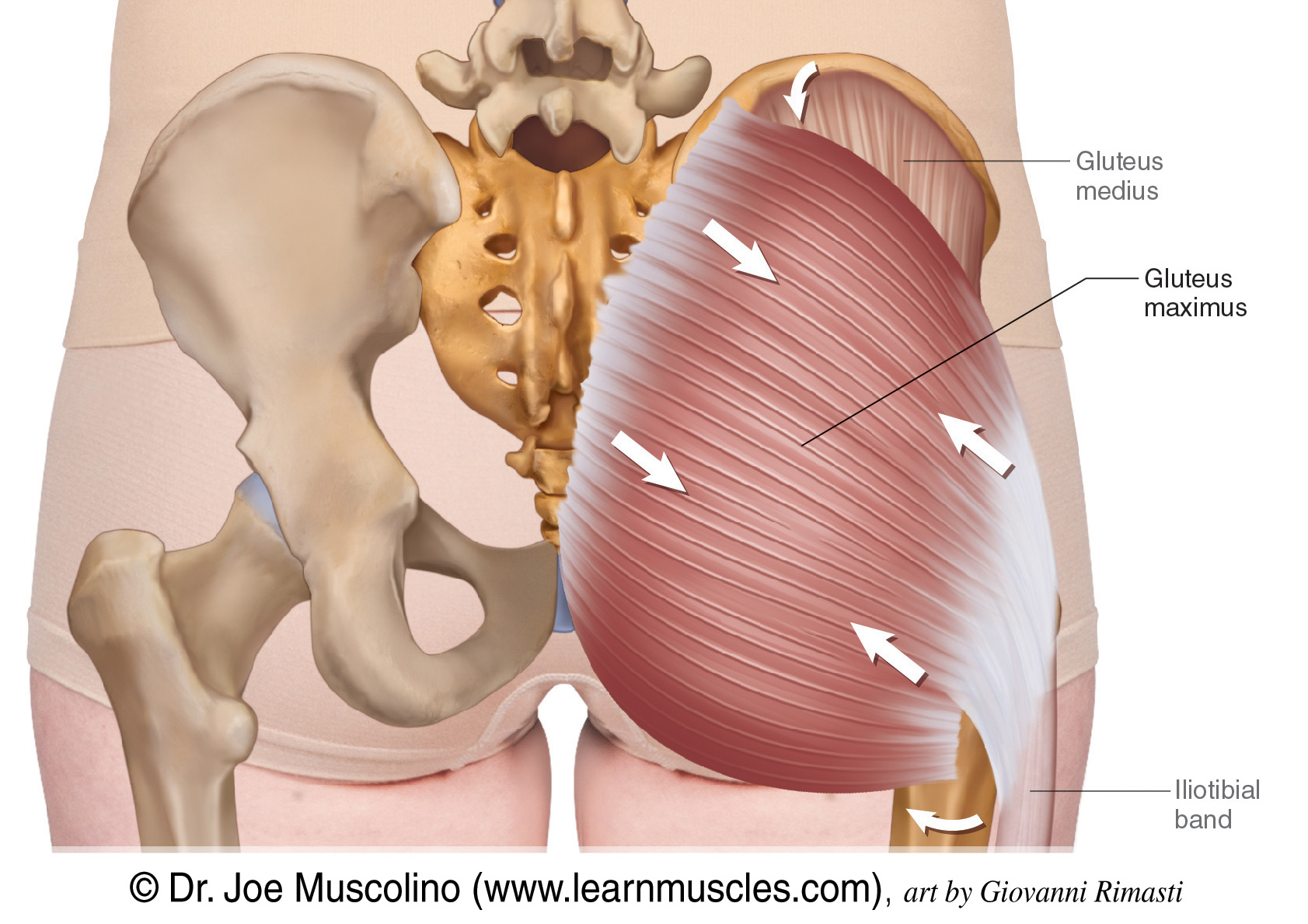
Gluteal Group Learn Muscles
Surface anatomy (also called superficial anatomy and visual anatomy) is the study of the external features of the body of an animal. [1] In birds, this is termed topography. Surface anatomy deals with anatomical features that can be studied by sight, without dissection.
The Glorious Glutes Muscles of the Buttocks
It also extends from the iliac crest superiorly to the gluteal fold inferiorly. The gluteal fold is the crease formed by the inferior aspect of the buttocks and the posterior upper thigh. Medially, the region extends to the mid-dorsal line and is called the intergluteal cleft, which is the groove that separates the buttocks from each other.
BUTTOCK MUSCLE BONES Anatomy Art Print for Medical Student Etsy
Surface anatomy of the buttocks. sus the posterior iliac crests, versus the ischia, versus the gluteal areas. Accurate identifi cation of wound location also infl uences the assessment of wound etiology. For example, terms "surface anatomy," "skin," "buttocks," and "gluteal suspicion of a pressure ulcer is heightened when it occurs area."

4 Anatomy of the buttocks and pelvis (Image by Springer) Download Scientific Diagram
Definition The buttock refers to the rounded bulge in the lower part of the gluteal region. The inferior extent of the buttock is marked by a gluteal fold of skin below. Medially, an intergluteal cleft separates the two buttock s from each other, while laterally they are bounded by the hip regions.